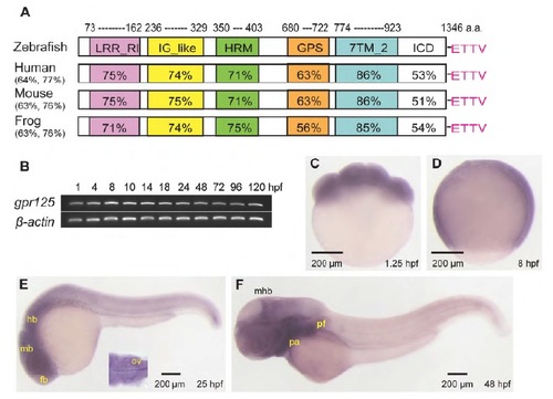
Predicted protein domains and spatiotemporal expression profile of gpr125 during early zebrafish development. (A) Schematics of predicted zebrafish Gpr125 protein domains. The percentage amino acid identity or similarity between Gpr125 peptide sequences in vertebrates for the whole protein are included in parentheses, and for individual domains on the schema. LRR_RI, leucine-rich repeat_ribonuclease inhibitor type; IG_like, immunoglobulin_like; HRM, hormone receptor domain; GPS, GPCR proteolytic site; 7TM_2, seven-pass transmembrane type 2; ICD, intracellular domain. The PDZ binding motif (ETTV) at the C-terminus are labeled in pink. (B) RT-PCR of gpr125 transcripts from 1 to 120 hpf. b-Actin was used as a loading control. (C-F) Whole-mount in situ hybridization profile of gpr125 expression. Lateral views, anterior is upwards in C,D and leftwards in E,F. fb, forebrain; hb, hindbrain; mb, midbrain; mhb, midbrain-hindbrain boundary; ov, otic vesicle; pa, pharyngeal arches; pf, pectoral fin.
|
