
Fig. 3
- ID
- ZDB-FIG-130815-15
- Publication
- Patterson et al., 2013 - Interactions with Iridophores and the Tissue Environment Required for Patterning Melanophores and Xanthophores during Zebrafish Adult Pigment Stripe Formation
- Other Figures
- All Figure Page
- Back to All Figure Page
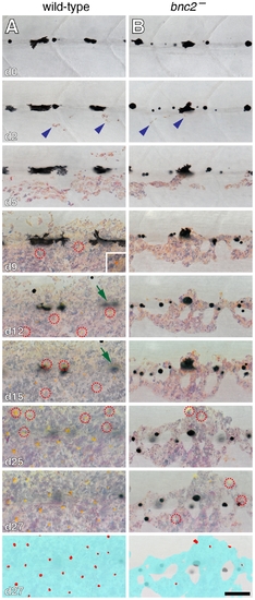
Interstripe xanthophores developed after iridophores in wild-type larvae and were further delayed in bnc2 mutants. Shown are a representative wild-type (bnc2/+) larva (A) and a sibling bnc2 mutant (B) imaged repeatedly over 27 d beginning at 6.0 SSL, just prior to the appearance of iridophores at the anteroposterior region imaged, dorsal to the anus. In both the wild-type and the bnc2 mutant iridophores started to appear by day 2 of imaging (blue arrowheads). Xanthophores started to differentiate by day 9 of imaging in wild-type; newly arising xanthophores are indicated by red dashed circles. In contrast, xanthophores did not appear until day 25 of imaging in the bnc2 mutant. As iridophores (and xanthophores) in the interstripe became more abundant, some early larval melanophores along the horizontal myoseptum disappeared from view (e.g., green arrows in A, d12 and d15). For easier visualization of melanophores and other cell type, fish were treated briefly with epinephrine immediately prior to imaging, which contracts melanosomes towards the cell body; the distribution of melanin thus indicates the centers of melanophores whereas processes extending out from the cell body are not visible. Bottom panels schematize the distribution of iridophores (light blue) and xanthophores (red) on the final day shown. Samples sizes for which complete image series were obtained were: bnc2, n = 4; bnc2/+, n = 6. Scale bar: in (B, d27) 80 μm for (A,B). |
| Fish: | |
|---|---|
| Observed In: | |
| Stage: | Days 30-44 |