
Fig. 1
- ID
- ZDB-FIG-130814-22
- Publication
- Pei et al., 2013 - Distinct Neuroblastoma-associated Alterations of PHOX2B Impair Sympathetic Neuronal Differentiation in Zebrafish Models
- Other Figures
- All Figure Page
- Back to All Figure Page
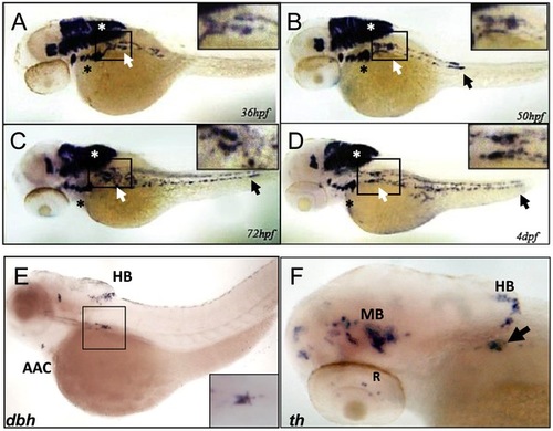
phox2b is expressed in the SCG, a marker of the peripheral sympathetic nervous system in the zebrafish. (A–D) Whole-mount in situ hybridization (ISH) for phox2b expression in wild-type embryos at the indicated time points. phox2b expression is seen in cells of the prospective superior cervical ganglion (SCG; white arrows, boxed) at 36 hpf (A), which start to extend caudally to form the sympathetic chain. These cells are identified as sympathetic neuronal cells by their expression of dbh and th (E, F). Expression is also seen in the brachial arches (black asterisk) and hindbrain (white asterisk). By 50 hpf, phox2b expression is seen in the enteric precursors (B–D, black arrow). Expression continues in two parallel rows caudally, encompassing the sympathetic chain until 4 dpf. (E) Lateral view (cranial to the left) of a 4-dpf zebrafish embryo analyzed by whole-mount ISH for dbh expression, which is seen in the SCG (black square), the arch associated complex (AAC) also derived from the neural crest, and the hindbrain (HB). Inset shows enlarged dorsal view of the SCG. (F) Whole-mount ISH of 4-dpf embryo analyzed for th expression. Aggregates of cells expressing th are seen in the SCG (arrow) as well as in the midbrain (MB), HB and retina (R). |
| Genes: | |
|---|---|
| Fish: | |
| Anatomical Terms: | |
| Stage Range: | Prim-25 to Day 4 |