
Fig. S2
- ID
- ZDB-FIG-130808-45
- Publication
- Jászai et al., 2013 - Spatial Distribution of Prominin-1 (CD133) - Positive Cells within Germinative Zones of the Vertebrate Brain
- Other Figures
- All Figure Page
- Back to All Figure Page
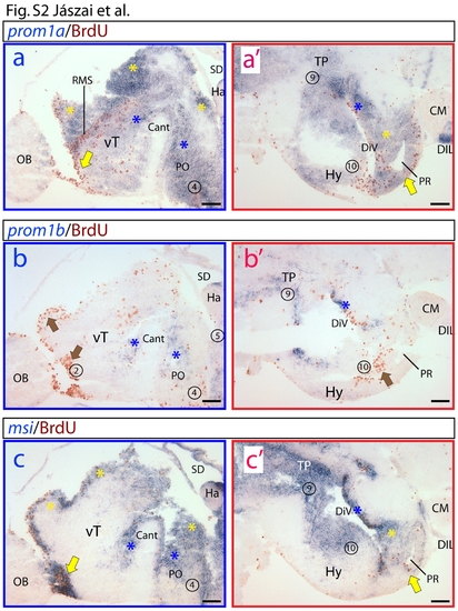
Differential expression of zebrafish prominin-1a and b in the prosencephalon. (a–c′) Higher magnification of insets displayed in panels A–C of Figure 6. Proliferative zones are indicated with numbers (see legend of figure 6). Coloured arrows and asterisks indicate overlapping expression domains of particular genes. (a, a′) Prominin-1a is strongly expressed in proliferating zones of the ventral telencephalon (vT) encompassing the rostral migratory stream-like assembly (RMS), preoptic area (PO), periventricular hypothalamus (PVH) including areas surrounding the posterior recessus (PR). Extending into the extraventricular, prominin-1a is detected in the diffuse nucleus of the hypothalamic inferior lobe (DIL). (b, b′) In contrast to prominin-1a, prominin-1b is excluded from the most extensively proliferating subdivisions of the prosencephalon (brown arrows). It is only weakly expressed in smaller subdomains of the vT, PO and PVH. (c, c′) Like prominin-1a, msi1 is detected in all of the extensively proliferating zones of the telencephalon (vT) and diencephalon (PO, PVH, posterior tuberculum (PT), PR). Cant, commissura anterior; CM, corpus mamillare; DiV, diencephalic ventricle; Ha, habenula; OB, bulbus olfactorius; SD, saccus dorsalis. Scale bars, a–c′, 100 μm. |
| Genes: | |
|---|---|
| Fish: | |
| Anatomical Terms: | |
| Stage: | Adult |