
- Title
-
Spatial Distribution of Prominin-1 (CD133) - Positive Cells within Germinative Zones of the Vertebrate Brain
- Authors
- Jászai, J., Graupner, S., Tanaka, E.M., Funk, R.H., Huttner, W.B., Brand, M., and Corbeil, D.
- Source
- Full text @ PLoS One
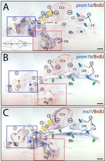
The combined expression of prominin-1a and b mimics the distribution of musashi-1 in adult zebrafish brain. (A–C) Cryosections of 3-month-old adult brain from BrdU-treated zebrafish were processed for ISH using an antisense DIG-labeled probe either against prominin-1a (A; prom1a), prominin-1b (B; prom1b) or musashi-1 (C; msi1). Proliferating cells were observed by immuno-detection of BrdU (brown). Proliferative zones are indicated with Arabic numbers (1 to 16, see below) from rostral to caudal direction along the neuraxis as proposed [41], and position of parasagittal section profiles of the brain is indicated on the cartoon (A) adapted from a standard neuroanatomical atlas of the zebrafish brain by Wulliman and colleagues [104]. The boxed areas in A–C are shown at higher magnification in Figure S2. Black dashed line indicates the border of prosencephalon towards the tegmentum of the brainstem (A). Coloured arrows and asterisks indicate overlapping expression domains of particular genes. Note that major sites of expression of prominin-1a are mainly located in the prosencephalic (rostral) and dorsal mesencephalic (tectal) domain (A), whereas prominin-1b is predominantly found in the rhombencephalic brainstem (caudal) domain (B). Msi1-1–positive cells are confined to all known proliferative zones both in rostral and caudal brain regions (C). CC, crista cerebellaris; CM, corpus mamillare; Cpost, commissura posterior; DIL, diffuse nucleus of the hypothalamic inferior lobe; Rh, rhombencephalon; SD, saccus dorsalis; T, telencephalon; 4V, fourth ventricle. Proliferative zones are: 1, olfactory bulb (OB); 2, ventral telencephalon; 3, dorsal telencephalon; 4, preoptic area; 5, ventral thalamus, 6, habenula; 7, periventricular zone of pretectum; 8, dorsal thalamus; 9, posterior tuberculum; 10, hypothalamus (Hy); 11, tectum opticum; 12, torus longitudinalis; 13, posterior mesencephalic lamina; 14a, molecular layer of valvula and corpus cerebelli (CCe); 14b, lobus caudalis cerebelli; 15, facial and vagal lobes; 16, ventricular zone of the fossa rhomboidea/canalis centralis. Zone 3 is indicated with a hollow number because is not represented on the present section. Black asterisk indicates rhombencephalic brain parenchyma. Scale bars, A–C, 250 μm. EXPRESSION / LABELING:
|
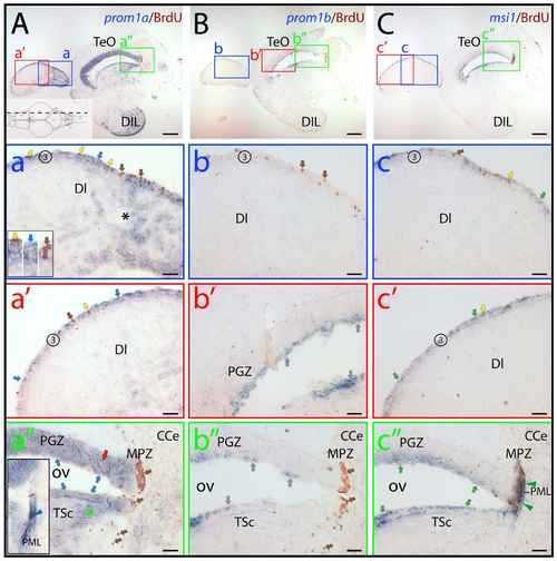
Distribution of zebrafish prominin-1a– and b–positive cells in the dorsal lateral telencephalon and the tectal ventricular zone. (A–C) Cryosections of 3-month-old adult brain from BrdU-treated zebrafish were processed for ISH using an antisense DIG-labeled probe either against prominin-1a (A; prom1a), prominin-1b (B; prom1b) or musashi-1 (C; msi1). Proliferating cells were observed by immuno-detection of BrdU (brown). Position of paramedian longitudinal sections of the brain is indicated on the cartoon (A) adapted from the neuroanatomical atlas by Wulliman and colleagues [104]. The boxed areas in A, B and C are displayed at a higher magnification in panels a–a′′, b–b′′ and c–c′′, respectively. (A) Prominin-1a–positive cells are enriched in the diffuse nucleus of the hypothalamic inferior lobe (A, DIL) and in the extraventricular dorsal telencephalic parenchyma (a, black asterisk). They are found within the proliferative zone 3 (3) of the dorsal telencephalic surface (a, a′, blue arrows), and are either BrdU–positive or negative (yellow and blue arrows, respectively, see inset in a). Rare proliferating cells are devoid of prominin-1a (a, brown arrow). Prominin-1a–positive cells are also detected in the superficial (blue arrows) and deeper (red arrow and green asterisk) areas of periventricular grey zone (PGZ) and Torus semicircularis (TSc) lining the optic (tectal) ventricle (OV) (a′′), respectively. While the marginal proliferating zone of the tectum (MPZ) lacks prominin-1a, a small number of prominin-1a–positive proliferating cells is observed in the posterior mesencephalic lamina (PML) (a′′, see inset, blue arrowhead). (B) Prominin-1b–positive cells (grey arrows) are found in the superficial tear of PGZ (b′) and in TSc lining the OV (b′′), whereas proliferating cells within the zone 3 of the dorsal telencephalic surface (b, brown arrows), the deeper areas of the PGZ (b′) and in the MPZ (b′′) are negative. (C) Msi-1–positive cells found within the proliferative zone 3 of the dorsal telencephalic surface (c, c′) are either BrdU–positive or negative (yellow and green arrows, respectively). Rare proliferating cells are devoid of msi-1 (c, brown arrow). Msi-1 is detected in the superficial tear of PGZ and TSc lining the OV (c′′) as well as in proliferating cells of the MPZ and those of the PML (c′′, green arrowheads). CCe, corpus cerebelli; DI, dorsal lateral subdivision of the dorsal telencephalic region; TeO, tectum opticum. Scale bars, A–C, 250 μm; a–c′′, 50 μm. EXPRESSION / LABELING:
|

Differential expression of zebrafish prominin-1a– and b–positive cells in the posterior dorsal telencephalon. (A–C) Cryosections of 3-month-old adult brain from BrdU-treated zebrafish were processed for ISH using an antisense DIG-labeled probe either against prominin-1a (A; prom1a), prominin-1b (B; prom1b) or musashi-1 (C; msi1). Proliferating cells were observed by immuno-detection of BrdU (brown). Position of the two coronal cross section profiles (I, II) encompassing the posterior dorsal telencephalic area is indicated on the cartoon adapted from a neuroanatomical atlas by Wulliman and colleagues [104], and the distribution of proliferative events (brown dots) is schematically shown. (A) Prominin-1a–positive cells are distributed along the posterior medial, dorsal and lateral aspects of the dorsal telencephalic surface proliferation zone (yellow arrows, see inset), and in the extraventricular brain parenchyma of the dorsal telencephalon (dT, asterisks) as well in the periventricular preoptic nucleus (PPa, blue arrows). (B) No prominin-1b–positive cells are detected within the dorsal telencephalic and preoptic proliferative zones (brown arrows). (C) Prosencephalic msi1–positive cells are found in a narrow ventricular stripe encompassing the proliferating subdivisions of the telencephalon, and that of the preoptic area and recessus opticus (Ro) of the diencephalic ventricle (green arrows). Proliferative zones 3 and 4 correspond to dorsal telencephalon and preoptic area, respectively. CO, chiasma opticum. Scale bars, A–C, 250 μm. EXPRESSION / LABELING:
|
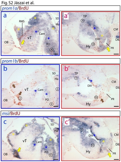
Differential expression of zebrafish prominin-1a and b in the prosencephalon. (a–c′) Higher magnification of insets displayed in panels A–C of Figure 6. Proliferative zones are indicated with numbers (see legend of figure 6). Coloured arrows and asterisks indicate overlapping expression domains of particular genes. (a, a′) Prominin-1a is strongly expressed in proliferating zones of the ventral telencephalon (vT) encompassing the rostral migratory stream-like assembly (RMS), preoptic area (PO), periventricular hypothalamus (PVH) including areas surrounding the posterior recessus (PR). Extending into the extraventricular, prominin-1a is detected in the diffuse nucleus of the hypothalamic inferior lobe (DIL). (b, b′) In contrast to prominin-1a, prominin-1b is excluded from the most extensively proliferating subdivisions of the prosencephalon (brown arrows). It is only weakly expressed in smaller subdomains of the vT, PO and PVH. (c, c′) Like prominin-1a, msi1 is detected in all of the extensively proliferating zones of the telencephalon (vT) and diencephalon (PO, PVH, posterior tuberculum (PT), PR). Cant, commissura anterior; CM, corpus mamillare; DiV, diencephalic ventricle; Ha, habenula; OB, bulbus olfactorius; SD, saccus dorsalis. Scale bars, a–c′, 100 μm. EXPRESSION / LABELING:
|

The combined expression of prominin-1a and b mimics distribution of musashi-1 in adult zebrafish brain. (A–C) Cryosections of 3-month-old adult brain from BrdU-treated zebrafish were processed for ISH using an antisense DIG-labelled probe either against prominin-1a (A; prom1a), prominin-1b (B; prom1b) or musashi-1 (C; msi1). Proliferating cells were observed by immuno-detection of BrdU (brown). Position of paramedian sections of the brain is indicated on the cartoon (A) adapted from a standard neuroanatomical atlas by Wulliman and colleagues [104]. Coloured arrows indicate overlapping expression domains of particular genes. Black dashed line indicates the border of prosencephalon towards the tegmentum of the brainstem (A). Major sites of prominin-1a (A) expression are located in the prosencephalic and tectal domains overlapping partly with msi1 (C) in certain subdivisions of the dorsal telencephalic surface proliferative zone (3), in a subdivision of the preoptic area (PO) located above the optic tract (OT), in the periventricular hypothalamus (PVH), in the periventricular grey zone (PGZ) and torus longitudinalis (TL). Expression of prominin-1a extends laterally into msi1–negative areas including the extraventricular parenchyma of the dorsal telencephalon (dT, asterisk), central parts of the ventral telencephalic area (Vc) and diffuse nucleus of the hypothalamic inferior lobe (DIL). Prominin-1b (B) overlaps with msi1 (C) in the periventricular rhombencephalon (griseum centrale, GC) and valvula cerebelli (Va). All three genes are detected in the dorsal thalamus (DT) and in the facial lobe (LVII). CC, crista cerebellaris; CCe, Corpus cerebelli; CM, corpus mamillare; Ha, habenula; LX, vagal lobe; OB, olfactory bulb; PR, posterior recessus of the diencephalic ventricle; VT, ventral thalamus. Scale bars, A–C, 250 μm. EXPRESSION / LABELING:
|