
Fig. 1
- ID
- ZDB-FIG-130807-3
- Publication
- Fang et al., 2013 - Characterization of transgenic zebrafish lines that express GFP in the retina, pineal gland, olfactory bulb, hatching gland, and optic tectum
- Other Figures
- All Figure Page
- Back to All Figure Page
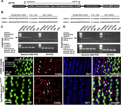
Crb2b expression in photoreceptors is not suppressed in transgenic fish Tg(LCRRH2-RH2-2:GFP)pt115. (A) A schematic illustrates the structures and tandem arrangement of a crb2b-targeting shRNA gene and a GFP reporter gene in the transgenic constructs for generating the Tg(RH2-2:GFP)pt115 fish lines. The region amplified in the PCR genotyping assay (see panel C) is indicated with a double-headed arrow. (B) The sequences of the sense and anti-sense regions of the crb2b-targeting shRNA genes are presented and the mismatching nucleotides are underlined. (C and D) PCR analyses suggest that the shRNA transgenes are present in all stable pt115 lines (arrow) at 5 dpf (C) and 9 mpf (D). However, RT-PCR analyses did not suggest apparent shRNA transgene transcripts at the size of genomic DNA PCR product. Genomic DNA PCR and RT-PCR of β-actin gene were conducted as controls for the quality of PCR DNA template, especially for genomic DNA contamination of cDNA. (E) The expression levels of Crb2b (red) in the transgenically expressing (arrowheads) and transgenically non-expressing retinal regions (arrows) were similar in adult shRNA-1 founder fish, suggesting no suppression of Crb2b by the shRNA transgenes. Transgenic cells were visualized by GFP reporter signals. Green cones are labeled with the letter G, and blue cones are labeled with letter B. The identification of cone types was based on the stereotypical spatial alignment of zebrafish cones in the order of UV–green–red–blue–red–green within the two dimensional photoreceptor mosaics ( Robinson et al., 1993, Raymond et al., 1995 and Zou et al., 2012) and green/red double cone (DC) staining with the zpr1 antibody. This identification method was also applied to Fig. 3, Fig. 4, Fig. 5 and Fig. 6. (F) In homozygous adult transgenic Tg(LCRRH2-RH2-2:GFP)pt11-k5 fish, the expression of Crb2b did not show apparent reduction because the expression levels at cone inner segment junctions next to transgenically-expressing cells (arrows) and non-transgenically-expressing cells (arrowheads) were identical. |
Reprinted from Gene expression patterns : GEP, 13(5-6), Fang, W., Bonaffini, S., Zou, J., Wang, X., Zhang, C., Tsujimura, T., Kawamura, S., and Wei, X., Characterization of transgenic zebrafish lines that express GFP in the retina, pineal gland, olfactory bulb, hatching gland, and optic tectum, 150-9, Copyright (2013) with permission from Elsevier. Full text @ Gene Expr. Patterns