
FIGURE
Fig. 2
- ID
- ZDB-FIG-130802-32
- Publication
- Collin et al., 2013 - ZNF408 is mutated in familial exudative vitreoretinopathy and is crucial for the development of zebrafish retinal vasculature
- Other Figures
- All Figure Page
- Back to All Figure Page
Fig. 2
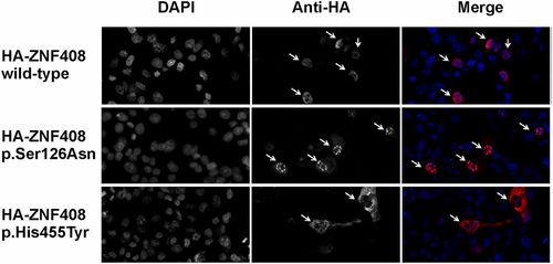
Immunocytochemical analysis of COS-1 cells transiently transfected with constructs encoding HA-tagged WT and mutant (p.Ser126Asn, p.His455Tyr) ZNF408 proteins. DAPI stains cell nuclei, whereas the anti-HA antibody stains the ZNF408 fusion proteins (indicated by arrows). (Right) Merged pictures (DAPI in blue, and HA-tagged ZNF408 proteins in red). Representative examples are shown for each transfection, indicating full nuclear localization for the WT and p.Ser126Asn ZNF408 proteins, but with the p.His455Tyr mutant ZNF408 confined mainly to the cytoplasm (each indicated by arrows). |
Expression Data
Expression Detail
Antibody Labeling
Phenotype Data
Phenotype Detail
Acknowledgments
This image is the copyrighted work of the attributed author or publisher, and
ZFIN has permission only to display this image to its users.
Additional permissions should be obtained from the applicable author or publisher of the image.
Full text @ Proc. Natl. Acad. Sci. USA