
Fig. S1
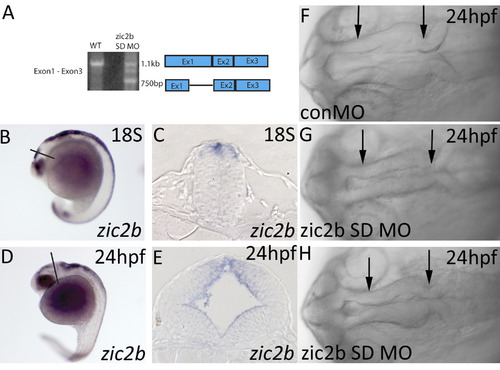
Zic2b expression and knockdown. (A) Embryos injected with zic2b splice-blocking MO express reduced levels of full-length zic2b transcript, and several mis-spliced transcript variants. (B,D) Wild-type embryos stained by ISH show expression of zic2b in the diencephalon, midbrain and hindbrain. (C,E) Restriction of zic2b expression to the dorsal neural tube is seen in transverse sections through the midbrain. (F–H) A midbrain neurulation defect is observed in zic2b morphants (midbrain primordium located between arrows in F–H). Zic2b morphants develop with small (35/104, 6 exp., see 1H) and closed midbrain ventricles by 24 hpf (33/104, 6 exp., see 1G). B,D are lateral views, anterior to the left. C,E are transverse sections through the midbrain. F–H are dorsal views, anterior to the left. |
Reprinted from Developmental Biology, 380(1), Teslaa, J.J., Keller, A.N., Nyholm, M.K., and Grinblat, Y., Zebrafish Zic2a and Zic2b regulate neural crest and craniofacial development, 73-86, Copyright (2013) with permission from Elsevier. Full text @ Dev. Biol.