
FIGURE
Fig. 2
- ID
- ZDB-FIG-130625-45
- Publication
- Lu et al., 2013 - A Novel Anti-Tumor Inhibitor Identified by Virtual Screen with PLK1 Structure and Zebrafish Assay
- Other Figures
- All Figure Page
- Back to All Figure Page
Fig. 2
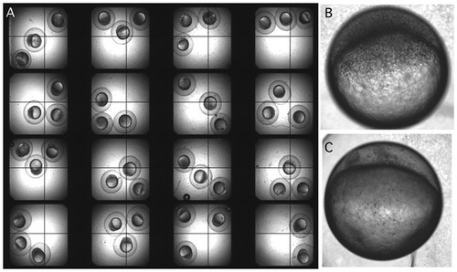
Screening for mitotic inhibitor using zebrafish embryo. p>A. Embryos were incubated with compounds in multi well plates and visualized under a dissection microscope. A compound was considered as positive if division of all three embryos in the well was inhibited. B. A normal embryo after 4 hours of development. C. An embryo with cell division inhibited at 1 cell stage when an active inhibitor was present even after 4 hours of development. |
Expression Data
Expression Detail
Antibody Labeling
Phenotype Data
Phenotype Detail
Acknowledgments
This image is the copyrighted work of the attributed author or publisher, and
ZFIN has permission only to display this image to its users.
Additional permissions should be obtained from the applicable author or publisher of the image.
Full text @ PLoS One