
Fig. 6
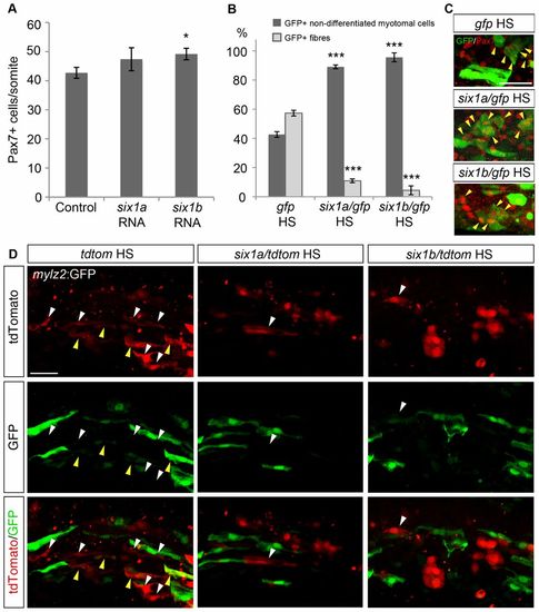
Overexpression of six1a and six1b promotes Pax7+ cell proliferation and inhibits fibre differentiation. (A) Average number of Pax7+ cells per somite in control embryos (n = 18) and embryos injected with six1a (n = 19) or six1b RNA (n = 17) at 24hpf. Control embryos have 42.7 Pax7+ cells/somite whereas the number is increased to 47.4 and 49.2 Pax7+ cells/somite after overexpressing six1a and six1b RNA, respectively. (B) Proportions of GFP+ fibres and GFP+ non-differentiated myotomal cells in 18-ss embryos after injecting a heat-shock-inducible construct driving the expression of gfp, either alone or together with six1a or six1b coding sequences, induced at 70% epiboly. The control GFP construct has 42.7% non-differentiated myotomal cells and 57.3% fibres (n = 6); induction of six1a expression results in 89.0% non-differentiated myotomal cells and only 11.0% fibres (n = 4); six1b overexpression gives a proportion of 95.6% non-differentiated myotomal cells and 4.4% fibres (n = 4). Somite number 8–10 was counted. (C) Expression of GFP (green) and Pax7 (red) in 18-ss embryos after inducing expression of gfp alone or together with six1a or six1b coding sequences at 70% epiboly. Yellow arrowheads indicate GFP+/Pax7+ cells. (D) Expression of GFP and tdTomato at 16ss in fast-fibre-specific myosin light chain 2:GFP, tg(mylz2:GFP)i135 embryos injected with a heat-shock-inducible construct driving the expression of tdtomato alone or together with six1a or six1b coding sequences, induced at 70% epiboly. Overexpressed six1a or six1b both resulted in a decreased occurrence of fibre-like morphology in the induced cells. Yellow arrowheads indicate GFP+/tdTomato+ fibres, white arrowheads indicate fibres only expressing tdTomato. *P<0.05, ***P<0.001. Scale bars: 50μm. |