FIGURE

Fig. 3

ID
ZDB-FIG-130618-18
Publication
Villefranc et al., 2013 - A truncation allele in vascular endothelial growth factor c reveals distinct modes of signaling during lymphatic and vascular development
Other Figures
All Figure Page
Back to All Figure Page
Fig. 3

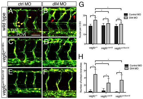
vegfcum18 partially rescues loss of dll4. (A-F) Two-photon micrographs of trunk blood vessels in fixed Tg(fli1a:egfp)y1 zebrafish embryos at 48 hpf immunostained for Fli1b. Anterior is to the left, dorsal is up. Yellow arrowheads indicate endothelial nuclei. Red arrows indicate ectopic vessel branches. Wild-type (A,B), vegfcum18/+ (C,D) and vegfcum18/um18 (E,F) embryos were injected with 15 ng control MO (A,C,E) or 15 ng Dll4 MO (B,D,F). (G,H) Quantification of (G) nuclei and (H) ectopic branches across three ISVs in MO-injected embryos of the indicated genotype. Values are the average of three experiments. *P<0.05; N.S., not significant; error bars indicate s.e.m. DA, dorsal aorta; PCV, posterior cardinal vein. Scale bar: 50 μm.

Expression Data
Gene:
Antibody:
Fish:
Knockdown Reagent:
Anatomical Terms:
Stage: Long-pec

Expression Detail
Antibody Labeling
Phenotype Data
Fish:
Knockdown Reagent:
Observed In:
Stage: Long-pec

Phenotype Detail
Acknowledgments
This image is the copyrighted work of the attributed author or publisher, and ZFIN has permission only to display this image to its users. Additional permissions should be obtained from the applicable author or publisher of the image. Full text @ Development