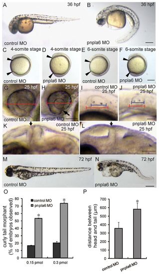
Knockdown of pnpla6 leads to distinct morphological change. (A) 36-hpf embryo injected with control MO. (B) 36-hpf pnpla6 morphant displays a curly tail. (C-F) Distinct shorter axis is seen in zebrafish embryos injected with pnpla6 MO: four-somite control MO embryo (C), four-somite pnpla6 MO morphant (D), six-somite control MO embryo (E) and six-somite pnpla6 MO morphant (F). (G,H) Eye development in control (G) and morphant (H) embryo with unclosed eye fissures (black arrow). (I,J) Otic vesicle development in control (I) and morphant (J) embryos. (K,L) Analysis of 25-hpf MHB defects, with the arrow denoting the location of the MHB: lateral view of MHB in control (K) and pnpla6 MO (L) embryos. (M) 72-hpf embryo injected with control MO. (N) 72-hpf pnpla6 morphant displays a swelling of the pericardium. (O) The dose-dependent effect on curly tails; *P<0.01 compared with control MO group. (P) The average distance between head and tail (straight-line length between two arrowheads) of early-stage (four-somite period) zebrafish embryos; *P<0.01 compared with control MO group. Scale bars: 200 μm (A-F); 50 μm (G,H); 100 μm (I,J); 50 μm (K,L); 200 μm (M,N).
|
