
Fig. 2
- ID
- ZDB-FIG-130510-9
- Publication
- Münch et al., 2013 - Notch regulates blastema proliferation and prevents differentiation during adult zebrafish fin regeneration
- Other Figures
- All Figure Page
- Back to All Figure Page
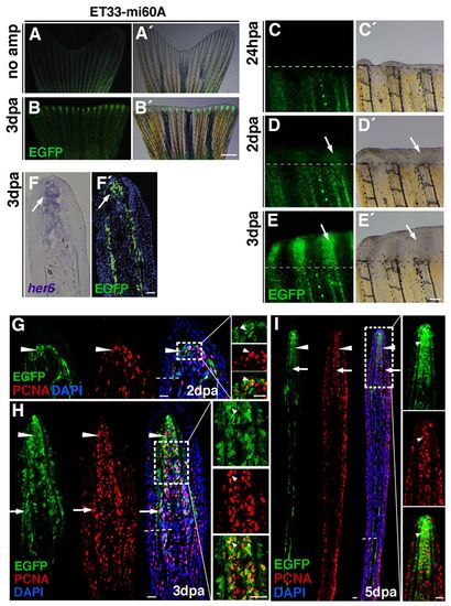
Lunatic fringe-mediated Notch signalling in proliferating blastema cells. (A-B2) EGFP expression in the enhancer trap line ET33mi60 is weak in non-amputated fins (A,A2), but strong in the fin regenerate at 3 dpa (B,B2). (C-E2) Time-course of ET-33mi60A regenerating fins. No EGFP expression is seen in the wound epidermis at 1 dpa. (C,C2). EGFP expression starts at 2 dpa within the blastema (D,D2, arrows). EGFP expression is strong within the blastema at 3 dpa (E,E2, arrows). (F,F2) her6 in situ hybridization and EGFP immunohistochemistry on a fin at 3 dpa reveals EGFP expression in cells in which Notch is activated (arrows). (G-I) EGFP and PCNA double immunohistochemistry. (G,H) Confocal microscopy images: EGFP is expressed during blastema formation (2 dpa) in cells co-expressing PCNA (G, arrowheads). At 3 dpa, EGFP expression is strong in all blastema cells (H, arrowheads), and is mosaic in proximal regions (H, arrows). Most cells are co-labelled with PCNA (H,I, arrowheads). At 5 dpa, EGFP expression is strong in the blastema (I, arrowheads) but weak in the proximal region (I, arrows). Most cells in the EGFP+ area express PCNA (I, arrowheads). Scale bars: 1 mm in B2 100 μm in E; 10 μm in E2,I. Broken lines mark the amputation plane. |
| Genes: | |
|---|---|
| Fish: | |
| Condition: | |
| Anatomical Terms: | |
| Stage: | Adult |