
Fig. S3
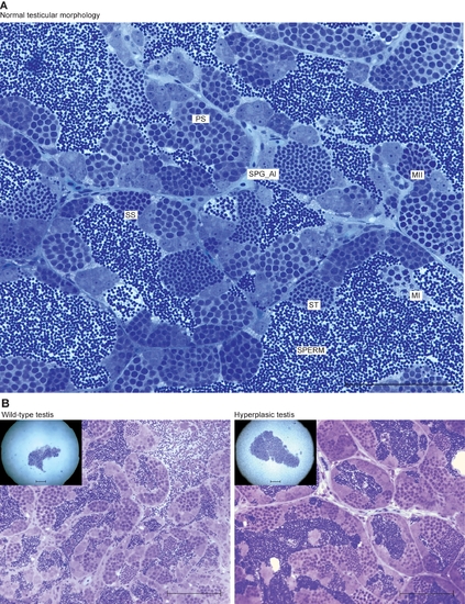
Zebrafish spermatogenesis and lrrc50Hu255h testicular hyperplasia. (A) The various stages of spermatogenesis in zebrafish can be morphologically distinguished and are indicated in a section from wild type zebrafish. Spermatogonial stem cells (SPG) that commit to spermatogenesis from clusters of paired (SPG_paired) and aligned spermatogonia (SPG_al) by mitotic divisions. All differentiated germ cells remain connected via stabilised intracellular bridges that allow the shared use of cytoplasmic components. This elegant mechanism is essential to synchronise collective mitosis, meiosis, differentiation and apoptosis of these clusters of cells. Next, SPG_al collectively differentiate into primary spermatocytes (PS), reducing cytoplasmic volume and severely modifying nuclear structure. Meiosis is the next step of differentiation, and occurs via the first (MI) and second (MII) meiotic divisions, forming haploid cells, these cells are known as secondary spermatocytes (SS). These cells contain one copy of the genome and need no further divisions; instead, these cells differentiate into spermatids (ST) that eventually form mature sperm. Scale bars; 100 μm. (B) Wild-type and hyperplastic testes of heterozygote lrrc50Hu255h zebrafish. A total of three have been identified, all showing moderate to extreme increases in testes volume, however upon histology we do not observe the typical increase in early germ cells as observed in tumors. Scale bars; 50 μm. |
| Fish: | |
|---|---|
| Observed In: | |
| Stage: | Adult |