
Fig. 6, S1
- ID
- ZDB-FIG-130502-45
- Publication
- Saxena et al., 2013 - Sox10-dependent neural crest origin of olfactory microvillous neurons in zebrafish
- Other Figures
- All Figure Page
- Back to All Figure Page
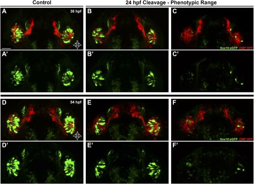
Shown are more details of the phenotype in Figure 6: (A and A′) control (antisense morpholino + sense photo-morpholino injected but not photocleaved) live embryos have robust numbers of ciliated (red) and neural crest–derived microvillous (green) neurons at 38 hpf. In contrast, identically injected embryos subjected to photocleavage at 24 hpf go on to develop ciliated neurons but have slight (B and B′) or, much more commonly, large (C and C′) decreases in microvillous neuron numbers and organization in comparison to control embryos. Photocleavage may have been less efficient in some embryos, resulting in a range of phenotypic severity. (D–F′) similar results are seen in the same embryos at 54 hpf, now with an even greater difference in microvillous neuron numbers between control and cleaved embryos. Sox10:eGFP: green; OMP:RFP: red. Orientation arrows: D: dorsal; V: ventral; L: lateral. Scale bars: 30 μm. |