|
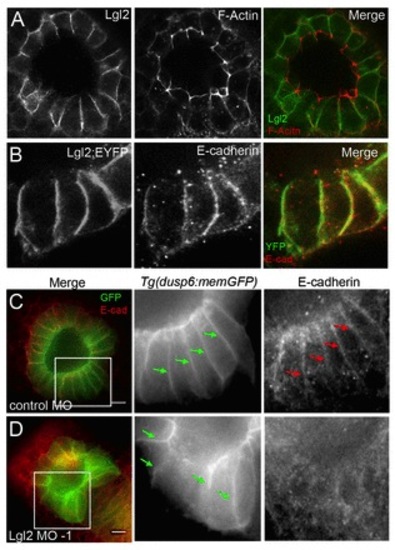
Lgl2 localizes to basolateral membranes in KV cells and E-cadherin is disrupted in Lgl2 knockdown embryos. (A) Fluorescence immunostaining using Lgl2 antibodies showed Lgl2 localized at basolateral membranes of KV cells and was excluded from the apical domain enriched with phalloidin staining of filamentous actin (F-Actin). (B) Lgl2:EYFP fusion protein also localized at basolateral membranes of KV cells and near E-cadherin staining. (C,D) In MO control Tg(dusp6:memGFP) embryos, E-cadherin localized at lateral membranes (C), whereas Lgl2 MO knockdown reduced membrane accumulation of E-cadherin (D). Boxes indicate enlarged regions shown in the individual channels. Arrows point out representative lateral membranes. Scale bars: 20 μm.
|