
Fig. 7
- ID
- ZDB-FIG-130412-14
- Publication
- Zhao et al., 2013 - The transcription factor Vox represses endoderm development by interacting with Casanova and Pou2
- Other Figures
- All Figure Page
- Back to All Figure Page
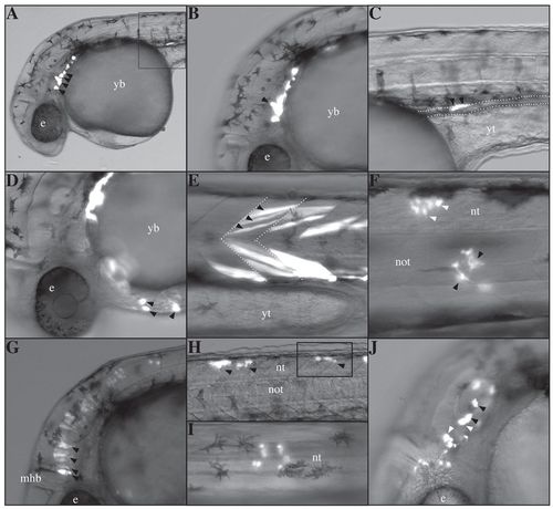
vox represses endodermal fates. Cells were grafted from donor sphere stage zebrafish embryos expressing either Tar*+GFP (A-D) or Tar*+vox+GFP RNAs (E-J) into host uninjected embryos from the same stage, which were allowed to develop and then examined by a combination of epifluorescence and brightfield microscopy at 30 hpf, except in F, which presents a 3-dpf embryo. Lateral views with anterior to the left, except for I which is a dorsal view. (A-D) The main fates induced by Tar* (a constitutively active form of taram-a) expression: pharyngeal endoderm (arrowheads in A,B, where the arrowhead in B points to the forming endodermal pharyngeal pouch), intestine (C, the inset from A; arrowheads points to two cells within the intestine, the contour of which is delineated by dotted lines) and the mesendodermal hatching gland (arrowheads in D). (E-J) The fates induced in Tar*+vox RNA-expressing cells: (E) trunk muscle cells (arrowheads) within the somites (delineated by dotted lines); (F) mesenchymal cells (surrounding the notochord, black arrowheads) and neural tube (white arrowheads); (G) hindbrain cells with their characteristic elongated shape; (H,I) posterior neural tube (arrowheads in H), I is a magnified dorsal view from the inset in H; (J) otic vesicle (black arrowheads) and epidermis (white arrowheads). e, eye; mhb, mid-hindbrain boundary; not, notochord; nt, neural tube; yb, yolk ball; yt, yolk tube. |