
Fig. 4
- ID
- ZDB-FIG-130321-10
- Publication
- Cheesman et al., 2011 - Epithelial cell proliferation in the developing zebrafish intestine is regulated by the Wnt pathway and microbial signaling via Myd88
- Other Figures
- All Figure Page
- Back to All Figure Page
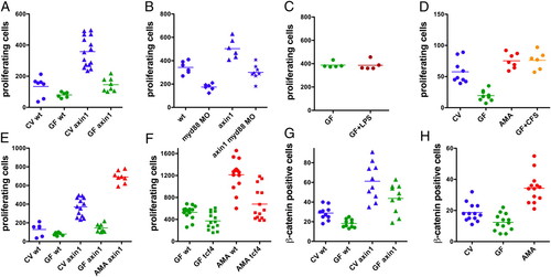
Combinatorial effects of microbial and Wnt signaling on intestinal epithelial cell proliferation. (A-F) S-phase intestinal epithelial cells were quantified in 30 serial sections in the intestinal bulb of individual larvae as in Fig. 1. To label S-phase nuclei, the experiments in A and D used BrdU and the experiments in B, C, E, and F used EdU. All experiments were performed with 6-dpf larvae except the experiment in D, which was performed with 8-dpf larvae. (A) CV axin1 had significantly more proliferating intestinal cells than CV WT (wt), GF wt, and GF axin1 (P < 0.0001). GF axin1 had significantly more proliferating cells than GF wt (P < 0.01). (B) axin1 had significantly more proliferating cells than wt, myd88 MO, and axin1 myd88 MO (P < 0.001). axin1 myd88 MO had significantly more proliferating cells than myd88 MO (P < 0.001). (C) Exposure of GF larvae from 3 to 6 dpf to 30 µg/mL LPS caused no change in cell proliferation relative to untreated GF larvae. (D) Monoassociation of GF larvae with A. veronii (AMA) or treatment with 500 ng/mL A. veronii CFS induced a significant increase in intestinal cell proliferation relative to GF (P < 0.0001). (E) axin1 mutants monoassociated with A. veronii had significantly more proliferating cells than CV axin1 (P < 0.0001). (F) tcf4 mutants had significantly fewer dividing intestinal cells than wt siblings when reared GF (P < 0.01) and when monoassociated with A. veronii (P < 0.0001). Intestinal epithelial cells with cytoplasmic β-catenin were quantified in 30 serial sections in the intestinal bulb of 6-dpf larvae. (G and H) CV wt larvae had significantly more β-catenin-positive cells than GF wt larvae (P < 0.05). (G) CV axin1 had significantly more β-catenin-positive cells than GF axin1 (P < 0.05). (H) A. veronii monoassociated larvae had significantly more β-catenin-positive cells than CV animals (P < 0.0001). |
| Fish: | |
|---|---|
| Conditions: | |
| Knockdown Reagent: | |
| Observed In: | |
| Stage: | Day 6 |