
Fig. 4
- ID
- ZDB-FIG-130318-7
- Publication
- Eisa-Beygi et al., 2013 - The 3-hydroxy-3-methylglutaryl-CoA reductase (HMGCR) pathway regulates developmental cerebral-vascular stability via prenylation-dependent signalling pathway
- Other Figures
- All Figure Page
- Back to All Figure Page
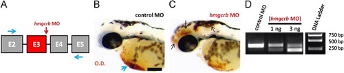
MO-mediated inhibition of the HMGCR pathway phenocopies ATV-treatment. (A) Partial hmgcrb pre-mRNA structure (not to scale) showing the exons (boxes), introns (black lines), the target of hmgcrb MO (red vertical arrow) and the positions of forward and reverse primers (black arrows) used to test the efficiency of the hmgcrb MO. ((B) and (C)) Representative bright-field micrographs of 48 hpf embryos injected with hmgcrb MO and control MO, stained with OD. Black arrows indicate sites of hemorrhage and blue arrow denotes the sinus venosus. Anterior is to the left and dorsal to the top. Scale bar=200 μm. (D) RT-PCR analysis of hmgcrb mRNA in hmgcrb morphant and control MO-injected embryos at 48 hpf. MO injection results in the dose-dependent appearance of a band corresponding to a transcript lacking exon-3. Doses of ATV are indicated above each lane. |
| Fish: | |
|---|---|
| Knockdown Reagent: | |
| Observed In: | |
| Stage: | Long-pec |
Reprinted from Developmental Biology, 373(2), Eisa-Beygi, S., Hatch, G., Noble, S., Ekker, M., and Moon, T.W., The 3-hydroxy-3-methylglutaryl-CoA reductase (HMGCR) pathway regulates developmental cerebral-vascular stability via prenylation-dependent signalling pathway, 258-266, Copyright (2013) with permission from Elsevier. Full text @ Dev. Biol.