
Fig. S6
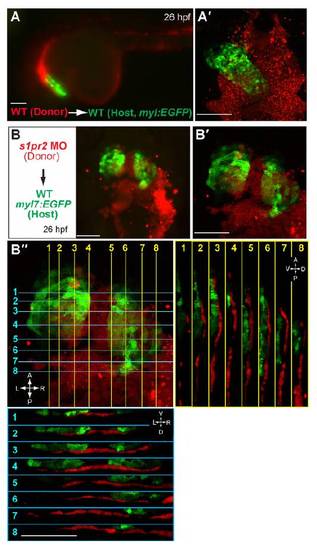
Donor cells do not contribute to myocardium when transplanted into endoderm. Epifluorescence images (A,B) and confocal z-stack images (A′,B′) showing rhodamin-dextran labeled donor cells and GFP-labeled myocardium in host embryos at 26 hpf. (A-A′) Wild-type donor cells were transplanted into wild-type hosts in the Tg(myl7:EGFP) background. (B-B′ ′) s1pr2 MO-injected donor cells were transplanted into wild-type hosts in the Tg(myl7:EGFP) background. (A) Lateral view; (A′′B,B′ ′ ventral view; (B′ ′) orthogonal view of B′ ′ Blue lines indicate cross-section planes along the L/R axis of the embryos perpendicular to the midline; yellow lines indicate cross-section planes along the embryonic AP axis; blue and yellow lines are numbered to show the position of the corresponding orthogonal panel; the respective sections are shown within blue or yellow insets. A, anterior; P, posterior; L, left; R, right; D, dorsal, V, ventral. Scale bars: 100 μm. |