|
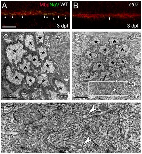
Nodes of Ranvier are disrupted in the spinal cord of st67 mutants and the spinal cord axons are hypomyelinated. (A,B) Myelin basic protein (Mbp, red) and NaV (green) antibody staining in the ventral spinal cord at 3 dpf. No differences can be seen in the intensity of Mbp stain in st67 mutants (B) compared with siblings (A), but NaV puncta (arrowheads) are greatly reduced in st67 mutants (see also Fig. 4). (C–E), TEM images showing cross-sections through the ventral spinal cord at 5 dpf. (C) At 5 dpf, many axons in sibling spinal cord are surrounded by several wraps of myelin (*). (D) Fewer axons are myelinated (*) in st67 mutant spinal cord, and irregular oligodendrocyte cytoplasm was observed with visibly swollen ER (arrowheads) in all mutants examined. (E) Enlarged view of the boxed region in D. In D and E, arrowheads denote swollen ER. Sample sizes: 8 wild-type and heterozygous larvae and 3 mutant larvae. Scale bars: 20 μm (A,B); 1 μm (C,D).
|