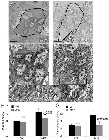
PLLn axons are hypomyelinated in st67 mutants. (A–E) TEM images showing cross-section through the PLLn at 5 dpf. Sorted axons(s) are completely surrounded by Schwann cells and some sorted axons are myelinated (*). In the wild type (A,C), many axons are surrounded by several wraps of myelin at 5 dpf. In st67 mutants (B,D), fewer axons are myelinated at 5 dpf, and irregular Schwann cell cytoplasm is observed with visibly swollen endoplasmic reticulum (D). (E) Blow-up of boxed region in D showing swollen ER (arrows). (F,G) A significant decrease in both sorted (F) and myelinated (G) axons was detected in st67 mutant larvae at 5 dpf, but not at 3 dpf. The P values for unpaired t-test comparisons (two-tailed) are shown; error bars indicate s.d. Sample sizes: at 3 dpf, 4 nerves from 4 siblings and 6 nerves from 5 mutants; at 5 dpf, 8 nerves from 5 siblings and 6 nerves from 3 mutants. All larvae were imaged at and quantifications made from approximately the same location along the anterioposterior axis, at the level of the 7th hemisegment. Scale bars: 2 μm (A,B); 0.5 μm (C,D).
|
