|
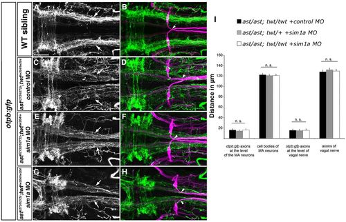
Medial displacement phenotype of otpb:gfp longitudinal HTS axons is not affected by depletion of sim1a in ast/ast; twt/+ and ast/ast; twt/twt embryos. Dorsal views of hindbrain confocal z-projections of 72 hpf otpb:gfp embryos. Anterior is towards the left. (A,B) otpb:gfp HTS axons (arrows) in a wild-type sibling. (C-H) Phenotypic strength of medial displacement of otpb:gfp-positive longitudinal axons is similar in ast/ast; twt/twt embryos injected with control MO (C, arrows), and in ast/ast; twt/+ (E, arrows) or ast/ast; twt/twt (G, arrows) sim1a morphant embryos. Arrowheads in B,D,F,H indicate pathfinding of Mauthner neurons. (I) Quantification of the distance between of otpb:gfp longitudinal axons and vagal axons, and of the distance between Mauthner neurons in ast/ast; twt/twt embryos injected with control MO, and in ast/ast; twt/+ or ast/ast; twt/twt embryos injected with sim1a MO. n.s., not significant. Scale bar: 50 μm.
|