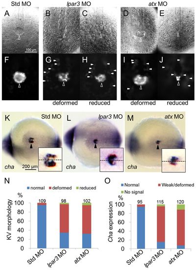
Kupffer′s vesicle formation is disturbed in both lpar3 and atx morphants. (A-J,N) Tg(sox17:dsRed) zebrafish embryos were injected with MO and the integrity of KV was examined under differential interference contrast (A-E) or epifluorescence (F-J) microscopy. In contrast to Std control MO-injected embryos (A,F), deformed (B,G,D,I) and/or reduced KVs (C,H,E,J) were observed in both lpar3 and atx morphants (open arrowheads). In addition, dispersed dsRed-positive cells (arrowheads) were also observed around KV in both lpar3 and atx morphants. To quantify KV defects, Tg(sox17:dsRed) embryos were used to repeat the above experiments and analyses are shown in N. (K-M,O) Embryos injected with the indicated MO were subjected to WISH against charon (cha, ventral view, bottom right). Arrowheads point to KVs. Magnified KV insets are shown at the lower right of each panel; embryo midlines are indicated by dashed lines. WISH against charon also revealed similar deformed/reduced pattern in lpar3 (L) and atx (M) morphants compared with Std control MO-injected embryos (K). Quantitative analyses are shown in O.
|
