
Fig. 3
- ID
- ZDB-FIG-130102-14
- Publication
- Kague et al., 2012 - Skeletogenic fate of zebrafish cranial and trunk neural crest
- Other Figures
- All Figure Page
- Back to All Figure Page
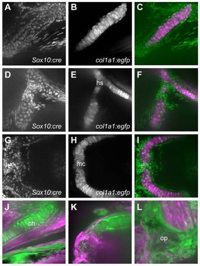
Chondrocytes and osteoblasts of the pharyngeal skeleton are NC-derived. A-I) Transgenics carrying a reporter that activates nuclear-Cherry expression following Cre activation (A, D, G) were crossed to -1.4col1a1:egfp transgenics, in which all cartilage cells are GFP+ (B, E, H). At 4 dpf, cells within the ceratohyal (A-C), hyosymplectic (D-F) and Meckel′s (G-I) cartilages have nucCh+ nuclei, indicating they are NC-derived. The GFP cells surrounding the cartilages, largely representing perichondral cells or osteoblast precursors, are also NC-derived. J-L) The reporter transgene switches from dsRed to GFP expression following Cre activation. At 10 dpf (J, K), cartilage cells of the ceratohyal (J) and Meckel′s (K) cartilages are GFP+, as are the cells surrounding them, indicating that the bone replacing the cartilages is also NC-derived. Bones forming via membranous ossification, such as the opercle (L), are also NC-derived. |
| Genes: | |
|---|---|
| Fish: | |
| Anatomical Terms: | |
| Stage Range: | Day 4 to Days 7-13 |