
Fig. 1
- ID
- ZDB-FIG-130102-12
- Publication
- Kague et al., 2012 - Skeletogenic fate of zebrafish cranial and trunk neural crest
- Other Figures
- All Figure Page
- Back to All Figure Page
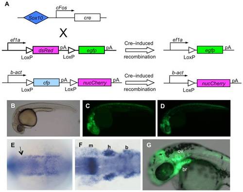
Cre recombinase permanently activates reporter gene expression in sox10 expressing cells of the zebrafish embryo. A) Diagram of transgenes used to genetically mark neural crest descendants; Cre activity under control of the Sox10 enhancer results in excision of the floxed first coding sequence in each reporter. In the first, dsRed is excised, leading to persistent expression of egfp under control of the ubiquitous ef1a promoter. In the second, cyan fluorescent protein (cfp) excision leads to persistent expression of nuclear mCherry (nucCh). B–D) At 24 hours post fertilization, egfp expression resulting from Cre activation (B, C) shows the same pattern as the expression under direct control of the Sox10 enhancer (D). E, F) Expression of cre is shown by in situ hybridization of a Sox10:egfp transgenic embryo. E) Early expression of cre is seen to the anterior extent of NC (arrow) flanking the neural keel at 6 somites. F) Expression persists in the mandibular (m), hyoid (h), and branchial (b) clusters of NC at 14 somites. G) At 30 hours, doubly transgenic embryos show robust expression of egfp in cells known to be derived from neural crest, including in the branchial arches (br), and in the otic vesicle (ov). B-D, G are side views with anterior to the left; E and F are dorsal views with anterior to the left. |
| Genes: | |
|---|---|
| Fish: | |
| Anatomical Terms: | |
| Stage Range: | 5-9 somites to Prim-15 |