FIGURE

Fig. S2

ID
ZDB-FIG-121212-2
Publication
Herbert et al., 2012 - Determination of Endothelial Stalk versus Tip Cell Potential during Angiogenesis by H2.0-like Homeobox-1
Other Figures
All Figure Page
Back to All Figure Page
Fig. S2

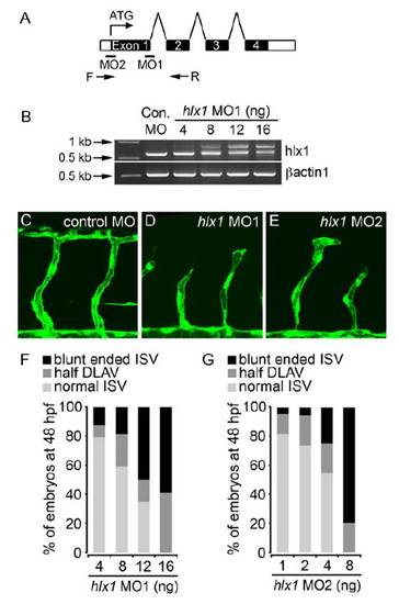
Knockdown of Hlx1 Using Gene-Targeted MO Reagents, Related to Figure 3
(A) Illustration of the hlx1 intron-exon structure indicating location of complementary PCR primers (arrows; F = forward, R= reverse) and MO-targeted sites. MO1 targets the first exon-intron boundary whereas MO2 targets the ATG start site.
(B) RT-PCR of hlx1 (using F and R PCR primers) or βactin1 using cDNA derived from embryos injected with either control MO or the indicated concentrations of MO1. MO1 dose-dependently alters hlx1 intron 1 splicing, resulting in a PCR product size shift.
(C-G) Lateral views of Tg(kdrl:GFP)s843 embryos at 48 hpf (C-E) and quantification of the morphology of individual ISVs (F, G; half DLAV = an ISV connected to only one adjacent ISV; blunt ended ISV = an ISV with no connections to adjacent ISVs) upon injection of embryos with either control MO or the indicated concentration of MO1 (D; 12 ng) or MO2 (E; 4 ng). Hlx1 knockdown disrupts ISV sprouting. Throughout this study hlx1 MOs were used as a combination of 4ng MO1 and 1ng MO2.

Expression Data

Expression Detail
Antibody Labeling
Phenotype Data

Phenotype Detail
Acknowledgments
This image is the copyrighted work of the attributed author or publisher, and ZFIN has permission only to display this image to its users. Additional permissions should be obtained from the applicable author or publisher of the image. Full text @ Curr. Biol.