
Fig. S5
- ID
- ZDB-FIG-121205-36
- Publication
- Garnett et al., 2012 - BMP, Wnt and FGF signals are integrated through evolutionarily conserved enhancers to achieve robust expression of Pax3 and Zic genes at the zebrafish neural plate border
- Other Figures
- All Figure Page
- Back to All Figure Page
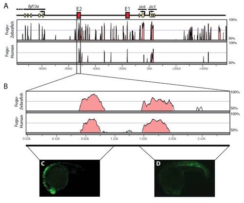
The neural plate border and mesodermal enhancer activities of zic3 E2 are separable. (A) The positions of zic3 E1 and E2 are shown with the level of sequence identity between Fugu and zebrafish and Fugu and human underneath. (B) The Vista plot comparing Fugu, zebrafish and human zic3 E2 regions indicates that there are two regions of high sequence identity within the enhancer. The 5′ and 3′ regions of zic3 E2 were separately placed upstream of the mouse Fos promoter and GFP, and their activities were analyzed in transiently transgenic embryos. (C,D) The 5′ region of E2 drives dorsal neural tube expression (C) and the 3′ region drives mesodermal GFP expression (D). C,D are lateral views with dorsal upwards and anterior towards the left. |