
Fig. 6
- ID
- ZDB-FIG-121119-7
- Publication
- Rodriguez-Mari et al., 2011 - Roles of brca2 (fancd1) in Oocyte Nuclear Architecture, Gametogenesis, Gonad Tumors, and Genome Stability in Zebrafish
- Other Figures
- All Figure Page
- Back to All Figure Page
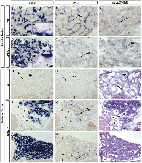
Altered Sertoli cell distribution, proliferation of spermatogonia, and accumulation of recombination-phase spermatocytes in brca2 mutant testes. In situ hybridization on adjacent sections of testes with markers for early germ cells (vasa), Sertoli cells (amh) and germ cells during recombination stages (sycp3) in wild types and brca2 mutants. (A-C) The anterior testis of wild types expressed vasa in early germ cells, amh in Sertoli cells surrounding tubules, and sycp3 in meiotic spermatocytes. (A′) Enlargement illustrates a gradient of vasa expression in spermatocytes: from moderate vasa expression (red circle) to low vasa expression (green circle), and finally spermatids and sperm (purple circle) that did not express vasa. (D) The anterior testis of brca2 mutants showed more tubules with vasa-expressing spermatogonia than wild types (A). (D′) Enlargement shows cells expressing low levels of vasa (red circle) and spermatocytes not expressing vasa (green circle) but no small spermatids or sperm. (E) brca2 mutants showed that amh-expressing cells were poorly organized and did not surround tubules neatly as in wild types, revealing an altered Sertoli cell distribution. (F) brca2 mutants expressed the recombination marker sycp3 in locally larger cell clusters than wild types, revealing an increased local concentration of cells at or entering pachytene stage. (G, H) In the posterior part of the testis, wild types did not express vasa or amh. (I) Hematoxylin and eosin (H&E) staining clearly showed sperm in many tubules in the posterior testes of wild types. (J-O) The posterior testes of brca2 mutants contained many tubules that were devoid of sperm (*) and contained vasa-expressing germ cells and Sertoli cells, which are not normally found in the wild-type posterior testes. (M-O) In one of two mutants analyzed, the posterior testes contained a larger proliferation of vasa-positive spermatogonia and also showed disorganized amh-expressing Sertoli cells, lacked sperm, and had empty tubules. Abbreviations: asterisks (*), empty testis tubules; gc, germ cells; i, intestine; ic, interstitial cells; sg, spermatogonia; sc, spermatocytes; Se, Sertoli cells; sp, sperm. |
| Genes: | |
|---|---|
| Fish: | |
| Anatomical Terms: | |
| Stage: | Adult |
| Fish: | |
|---|---|
| Observed In: | |
| Stage: | Adult |