
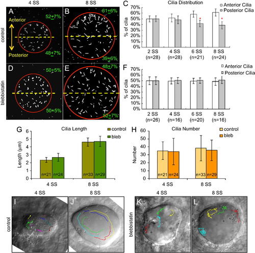
Fig. 5
Blebbistatin disrupts asymmetric cilia distribution and directional fluid flow in KV. (A, B, D and E) Immnostaining of KV cilia in control DMSO treated (A and B) and blebbistatin treated (D and E) embryos at 4 SS and 8 SS. (C and F) Analysis of cilia distribution in DMSO control (C) and blebbistatin (F) treated embryos during KV development. Error bars=one standard deviation. n=number of embryos analyzed. *Significant difference between anterior and posterior (p<0.0001). (G and H) KV cilia length (G) and number (H) was similar in control and blebbistatin treated embryos at 4 SS and 8 SS. Error bars=one standard deviation. n=number of embryos analyzed. (I–L) Visualization of fluid flow by superimposing tracks of bead movement on an image of KV (see Movies S4–S7). Control DMSO treated embryos displayed uncoordinated flow at 4 SS (I), but strong counter-clockwise flow at 8 SS (J). In blebbistatin treated embryos, directional flow was not observed at 4 SS (K) or 8 SS (L). |
Reprinted from Developmental Biology, 370(1), Wang, G., Manning, M.L., and Amack, J.D., Regional Cell Shape Changes Control Form and Function of Kupffer's Vesicle in the Zebrafish Embryo, 52-62, Copyright (2012) with permission from Elsevier. Full text @ Dev. Biol.