
Fig. S1
- ID
- ZDB-FIG-121101-28
- Publication
- Cox et al., 2012 - An essential role of variant histone h3.3 for ectomesenchyme potential of the cranial neural crest
- Other Figures
- All Figure Page
- Back to All Figure Page
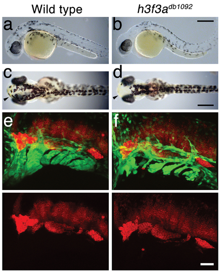
NC derivatives in h3f3adb1092 mutants. a–d, Live views at 32 hpf (a, b) and 54 hpf (c, d) show reductions of head melanocytes (black) in h3f3adb1092/db1092 homozygotes (n = 22/28). The melanocytes of the eye and trunk were never affected. Cranial xanthophores (yellow), most clearly seen at the anterior limit of the head (arrowheads), were also largely unaffected. e, f, In confocal projections of fli1a:GFP embryos at 36 hpf, HuC antibody staining (red) labels neurons of the cranial ganglia – from left to right the trigeminal, anterior lateral line, auditory, and posterior lateral line – which are unaffected in h3f3adb1092/db1092 homozygotes (n = 8). In the merged images, fli1a:GFP (green) shows a reduction of CNC-derived ectomesenchyme in the mutant. Scale bars: a–d, 250 μm; e & f, 50 μm. |