
Fig. 6
- ID
- ZDB-FIG-121031-6
- Publication
- Andersen et al., 2012 - Centrosome movements in vivo correlate with specific neurite formation downstream of LIM homeodomain transcription factor activity
- Other Figures
- All Figure Page
- Back to All Figure Page
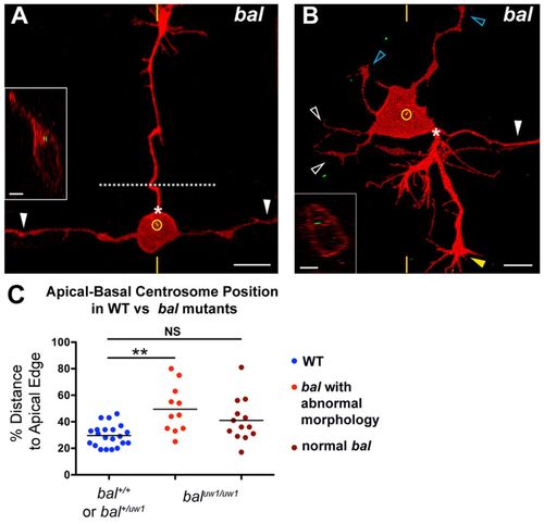
RB neurons have mislocalized centrosomes and aberrant morphology in bal mutant embryos. (A,B) Individual RB neurons labeled by transient expression of TagRFP-CAAX in GFP-Xcentrin mRNA-expressing bal mutant zebrafish embryos. Dorsal-lateral views, anterior left. Images are z-projections and insets are optical cross-sections through the centrosome region (indicated with yellow hatch marks). (A) Mature RB neuron in a bal embryo with normal central axons (white arrowheads) and an ectopic apical axon (PAS indicated by asterisk) that crossed the midline (dotted line) and grew to the contralateral trunk. The centrosome (yellow circle) is localized near the apical cell body edge. (B) Mature RB neuron in a bal embryo with supernumerary axons. Neuron extended a normal descending central axon (white arrowhead) and peripheral axon (yellow arrowhead) from the PAS (asterisk), but has truncated ascending central neurites (white open arrowheads) and ectopic apical neurites (blue open arrowheads). The centrosome (yellow circle) is localized halfway between the apical and basal cell surfaces. (C) Scatter plot of centrosome position. Mean ± s.e.m. for WT: 29.6±1.8 (n=21); abnormal bal, 49.5±5.4 (n=11); normal bal, 41.1±4.5. ANOVA with Tukey’s multiple comparison shows that abnormal bal is significantly different from WT (**P<0.05) and that normal bal is not significantly different (NS) from WT. |