
FIGURE
Fig. 1
- ID
- ZDB-FIG-121025-7
- Publication
- He et al., 2012 - How variable clones build an invariant retina
- Other Figures
- All Figure Page
- Back to All Figure Page
Fig. 1
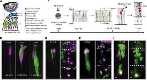
In Vivo Mosaic Labeling of Single RPCs (A) A schematic of the retina and its major cell types. (B) Experimental flow of labeling and tracking a retinal clone from a photoconverted single RPC (magenta). (C) Single RPCs (magenta) photoconverted at 24 hpf from the clones of various size (green). (D–F) Single RPCs photoconverted at 24 hpf (D), 32 hpf (E), and 48 hpf (F) (magenta, left) and resultant clones (magenta, right) with cell fates identified (Figure S1). Scale bar represents 5 μm. |
Expression Data
Expression Detail
Antibody Labeling
Phenotype Data
Phenotype Detail
Acknowledgments
This image is the copyrighted work of the attributed author or publisher, and
ZFIN has permission only to display this image to its users.
Additional permissions should be obtained from the applicable author or publisher of the image.
Full text @ Neuron