|
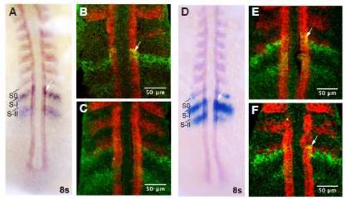
Expression of mesp-ab and mesp-bb in the anterior presomitic mesoderm. (A) Double in situ showing expression of myod1 (red) and mesp-ab (blue) at 8-somite stage. (B,C) Fluorescent double in situ showing expression of myod1 (red) and mesp-ab (green) at 8-somite stage. The most anterior stripe of mesp-ab in S-I is immediately adjacent to the caudal myod1 expression in S0. Dynamic mesp-ab expression also overlaps with myod1 expression in the adaxial cells (white arrows). (D) Double in situ showing expression of myod1 (purple) and mesp-bb (blue) at 8-somite stage. (E,F) Fluorescent double in situ showing expression of myod1 (red) and mesp-bb (green) at 8-somite stage. Expression of mesp-bb in S-I abuts myod1 expression in S0. Dynamic mesp-bb expression also overlaps with myod1 expression in the adaxial cells (white arrows).S0, S-I and S-II mark the position of presumptive somites. All embryos were flat-mounted and are shown in dorsal views, anterior to the top.
|