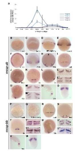
Embryonic expression of mesp-ab and mesp-bb. (A) qPCR time course of mesp gene expression from 1-cell to prim-5 (24 hours-postfertilization). Expression is shown in units relative to total 18S RNA expression at the stage shown. (B,C) Expression of mesp-ab is not detected at 1-cell or dome stage. Animal to the top. (D,E) mesp-ab expression initiates in the ventral and lateral margin at 40% epiboly and persists in a similar pattern at shield stage. Dorsal view, animal to the top. (F) Animal view of shield stage mesp-ab expression, ventral to the top. (G,H) As gastrulation proceeds and mesoderm ingresses, expression persists in the lateral mesoderm (arrows), and is down-regulated on the ventral side (arrowhead). (G) Shows dorsal view, animal to the top, (H) shows animal view, ventral to the top. (I) At the end of gastrulation stripes are seen in the paraxial mesoderm. Dorsal view, anterior to the top. (J) Anterior view, ventral to the top, of bud stage mesp-ab expression in paraxial meosderm. (K,O) mesp-ab expression persists in paraxial mesoderm during somitogenesis. (K) Dorsal view, anterior to the top, at 3-somite stage showing one stripe of expression. (L) Lateral view, anterior to the top left, at 3-somite stage showing one stripe of expression. (L′-L′′) Enlarged dorsal view of mesp-ab expression in paraxial mesoderm at 3-somite stage showing one or two stripes of expression (asterisks). (M) Lateral view, anterior to the top left, at 12-somite stage. (M′-M′′) Enlarged dorsal view of mesp-ab expression in paraxial mesoderm at 12-somite stage showing one or two stripes of expression (asterisks). (N) Lateral view, anterior to the left, of mesp-ab expression in the tail at 26 somite stage. (O) Dorsal view, anterior to the top, of mesp-ab expression in the tail at 26-somite stage. (P,Q) Expression of mesp-bb is not detected at 1-cell stage or throughout early development; 70% epiboly shown as an example. Animal to the top. (R) mesp-bb expression is first detected at bud stage when stripes are seen in the paraxial mesoderm. Dorsal view, animal to the top. (S) Animal view, ventral to the top, of bud stage mesp-bb expression in the paraxial mesoderm (arrows). (T-W) mesp-bb expression persists in paraxial mesoderm during somitogenesis.(T) Dorsal view, anterior to the top, at 3-somite stage showing two stripes of expression. (T′-T′′) Enlarged dorsal view of mesp-bb expression in paraxial mesoderm at 3-somite stage showing two or three stripes of expression (asterisks). (U) Lateral view, anterior to the top left, at 12-somite stage showing two stripes of expression. (U′-U′′) Enlarged dorsal view of mesp-bb expression in paraxial mesoderm at 8-12-somite stage showing one or two stripes of expression (asterisks). (V) Lateral view, anterior to the left, of mesp-bb expression in the tail at 26-somite stage. (W) Dorsal view, anterior to the top, of mesp-bb expression in the tail at 26-somite stage.
|
