
Fig. 4
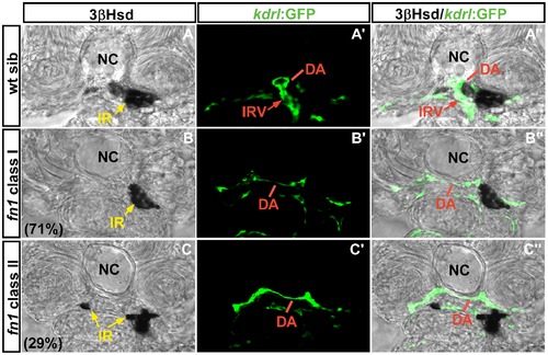
The interrenal tissue and peri-interrenal vasculature phenotypes in the fn1 mutant. Transverse sections of fnkt259;Tg(kdrl:EGFP)s843 embryos and their wild-type siblings at 3 dpf were visualized for 3β-Hsd activity (black) and GFP (green). All sections are oriented with the posterior end toward top of page. The images for wild-type siblings (A–A′′) were acquired by focusing on the sprouting point of the IRV from the DA, and hence its surrounding steroidogenic cells were masked by the endothelial structure on panel A. The IRV angiogenesis and interrenal morphogenetic movements are severely disrupted in the fn mutant. Yellow arrow, interrenal tissue (IR). Abbreviations: notochord (NC), interrenal tissue (IR), dorsal aorta (DA), interrenal vessel (IRV). |
| Gene: | |
|---|---|
| Fish: | |
| Anatomical Term: | |
| Stage: | Protruding-mouth |
| Fish: | |
|---|---|
| Observed In: | |
| Stage: | Protruding-mouth |