
Fig. 6
- ID
- ZDB-FIG-120814-38
- Publication
- Mitra et al., 2012 - Requirement for a uroplakin 3a-like protein in the development of zebrafish pronephric tubule epithelial cell function, morphogenesis, and polarity
- Other Figures
- All Figure Page
- Back to All Figure Page
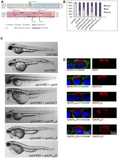
The CT of Upk3l is critical for its function. (A) Clustal W alignment of the TM and CT region of hUP3a, xUP3a and Upk3l. The amino acids that comprise the TM and CT domain are highlighted, and conserved functional motifs and residues are underlined or shaded, respectively. (B) Phenotypes of embryos injected with CNT-MO, upk3l-MO alone, or upk3l-MO co-injected with mCherry mRNA and mRNAs encoding MO-resistant versions of upk3lΔCT, upk3lP258L, upk3lY251F, or upk3lY251D. Microinjection of MO-resistant mRNA alone at the same doses did not produce detectable phenotypic abnormalities. (C) Morphological phenotypes associated with embryos injected with 5 ng CNT-MO (n = 100), 3 ng of upk3l-MO (n = 100), or 3 ng of upk3l-MO and 100 pg of upk3l, upk3lΔCT, upk3lP258L, upk3lY251F, or upk3lY251D mRNA (n≥50). (D) Localization of FLAG-tagged Upk3l, Upk3lΔCT, Upk3lP258L, Upk3lY251F, or Upk3lY251D in MDCK cells co-stained for actin and nuclei. Upk3lY251F and Upk3lY251D showed a predominantly intracellular localization and their low levels of expression necessitated an 1.5-fold increase in photomultiplier voltage above that used for the other samples. |