
Fig. S2
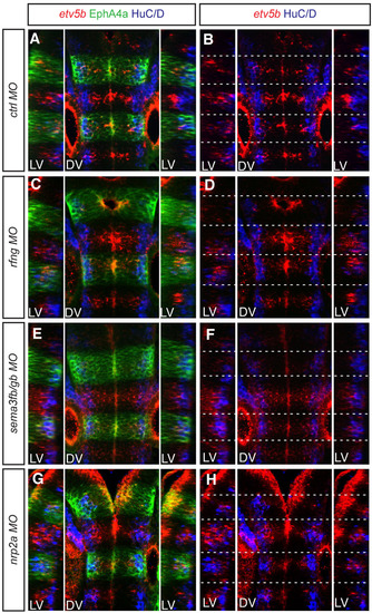
etv5b expression in rfng, sema3fb/gb and nrp2a morphants. Confocal images of dorsal views of the 30-hpf zebrafish hindbrain, anterior to the top, for (A,B) control MO, (C,D) rfng MO, (E,F) sema3fb+sema3gb MOs and (G,H) nrp2a MO. Dashed white lines indicate the position of segment borders. Left and right reconstructed lateral views (LV) plus dorsal views (DV) of etv5b whole-mount mRNA fluorescent in situ hybridisation (red), combined with HuC/D (blue) and EphA4 antibody staining (green). etv5b expression is less restricted to segment centres and/or is at lower levels in all the knockdown conditions compared with the control situation. Orientation of embryos as Fig. 1. |