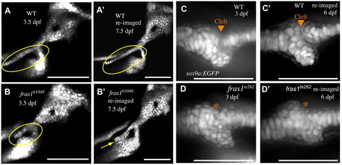
Me-Pq fusion occurs by 72 hpf, whereas Sy-Ch fusion often occurs later. (A-B′) Confocal sections of second arch-derived zebrafish skeleton at 3.5 dpf (A,B) and the same skeletal elements reimaged later in development (A′,B′). Anterior to left, dorsal up. By 3.5 dpf, a large gap is present between WT symplectic and ceratohyal cartilages (A), and this gap persists through larval development (A′). In fras1 mutants, although there is often a space between symplectic and ceratohyal cartilage (B, circled) at 3.5 dpf, these skeletal elements are typically found fused together when examined later in larval development (B′, yellow arrow). (C-D′) Confocal projections of first arch joint region, shown anterior to left and dorsal up. By 3 dpf, WT embryos have formed a cleft between Meckel′s and palatoquadrate cartilages (C), which persists as the embryos develop (C′). When fras1 mutants show skeletal fusions in the first arch (D, asterisk) they are always visible by 3 dpf, and persist when re-examined later in larval development (D′). Scale bars: 100 μm.
|
