
Fig. 5
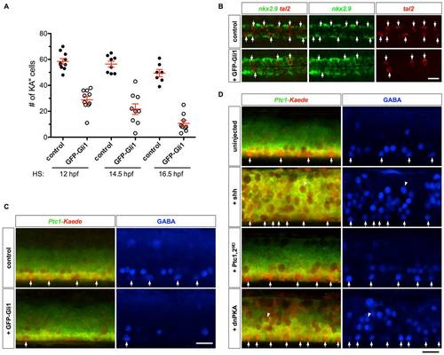
Prolonged Hh response interferes with KA′′ specification. (A) Quantification of KA3 specification in embryos overexpressing GFP-Gli1. hsp-GFP-Gli1 embryos and their non-transgenic sibling controls were heat-shocked at indicated stages, and stained at 25 hpf for the expression of tal2. (B) hsp-GFP-Gli1 and control embryos were heat-shocked at 14 hpf, and stained at 24 hpf for the expression of tal2 and nkx2.9. Induction of GFP-Gli1 results in a reduction of tal2-positive KA′′ cells (arrows) and expansion of nkx2.9-expressing LFPs. (C) Ptc1-Kaede control embryos, and Ptc1-Kaede; hsp-GFP-Gli1 embryos were heat-shocked at 14 hpf, photoconverted at 24 hpf, and stained with the GABA antibody (blue) at 36 hpf. Induced expression of GFP-Gli1 results in a reduction of KA′′ cells (arrows). Note that at 36 hpf, GFP-Gli1 expression has minimal contribution to the green fluorescence. (D) Ptc1-Kaede control embryos, and Ptc1-Kaede embryos injected with Shh mRNA, Ptc1 and Ptc2 morpholinos, or dnPKA mRNA were photoconverted at 24 hpf, and stained with the GABA antibody (blue) at 36 hpf. Arrows indicated GABA-positive cells in the LFP domain. Note that Shh and dnPKA overexpression induced many ectopic GABAergic neurons (arrowheads) throughout the dorsal-ventral axis of the spinal cord, and most of them appeared to lose Hh response by 24 hpf as indicated by the expression of Ptc1-Kaedered. Scale bars: 20 μm. |