
Fig. 6
- ID
- ZDB-FIG-120710-14
- Publication
- Soares et al., 2012 - Dre-miR-2188 Targets Nrp2a and Mediates Proper Intersegmental Vessel Development in Zebrafish Embryos
- Other Figures
- All Figure Page
- Back to All Figure Page
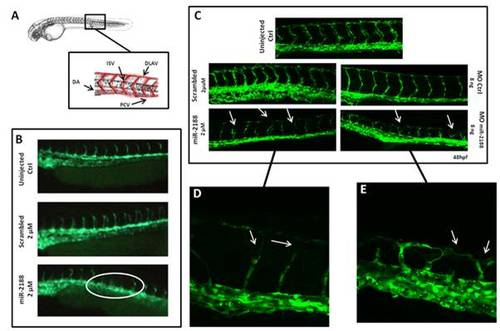
Analysis of blood vessel formation in Tg(flk1-GFP)s843 embryos. A) Representation of the zebrafish circulatory system showing the major structures (DLAV - Dorsal Longitudinal Anastomotic Vessel; ISV – Intersegmental Vessel; DA – Dorsal Aorta; PCV – Posterior Cardinal Vein). B) Visualization of 24 hpf embryo blood vessels using fluorescence microscopy. After miR-2188 duplex microinjection under developed and absent ISVs were observed, non injected and scrambled duplex injected embryos did not show such defects. C) Visualization of 48 hpf embryo blood vessels using confocal microscopy (20x). ISVs of miR-2188 injected embryos were thinner than those of non injected and scrambled duplex injected embryos and displayed ISV’s patterning defects (arrows). MOmiR-2188 injected embryos revealed DLAV defects and branching of ISVs (arrows). Images shown in D) and E) are 60x amplification images of miR-2188 duplex and miR-2188-MO injected embryos, respectively. |
| Fish: | |
|---|---|
| Knockdown Reagent: | |
| Observed In: | |
| Stage: | Long-pec |