
Fig. 1
- ID
- ZDB-FIG-120601-45
- Publication
- Stobb et al., 2012 - Graph theoretical model of a sensorimotor connectome in zebrafish
- Other Figures
- All Figure Page
- Back to All Figure Page
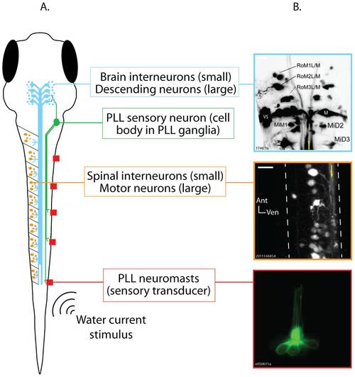
The posterior lateral line (PLL) sensorimotor pathway in zebrafish larvae. A. Schematic representation of a 6 day old zebrafish larva (dorsal view) showing, in different colors, all of the neuron types included in the model pathway, and the effective stimulus for hair cell activation (water current). Neuron types are labeled to the right of the schematic. B. Photomicrographs showing individually identifiable neurons within the PLL sensorimotor pathway. Top: Descending neurons in the hindbrain of a 6 day old zebrafish larva after injection labeling with a fluorescent tracer. The Mauthner neurons (M), Mauthner segmental homologs (Mid2, MiD3), and other identifiable descending neurons can be seen. The image is a maximum projection of 22 optical sections taken at 3 µm intervals through the dorsal-ventral axis of the hindbrain. Anterior is up and the yellow line shows the approximate midline. Middle: Spinal neurons imaged in a 7 day old larva prepared as described for A. The image is a maximum projection of 10 optical sections taken at 2 µm intervals through the rostral spinal cord at approximately the level of the 8th myotome. The dashed white lines trace the spinal cord′s boundaries, and the yellow arrow is drawn along the bundle of descending axons running down the ventral spinal cord. Bottom: A PLL neuromast. This neuromast was imaged in a single optical section in a 6 day old Brn3c:eGFP transgenic larva, in which all lateral line neuromasts express GFP. Individual hairs (cilia) can be seen extending toward the top of the image. Sensory neurons (not shown) contact the hair cell bodies, which can be seen at the bottom of the image. Scale bars = 20 μm. |
| Gene: | |
|---|---|
| Fish: | |
| Anatomical Term: | |
| Stage: | Day 6 |