
Fig. 1
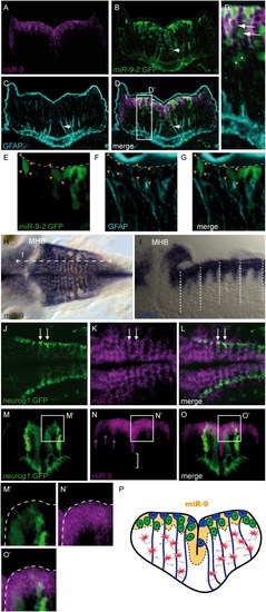
miR-9 Expression Encompasses Different Progenitor Commitment Stages across the Hindbrain Ventricular Zone (A–G) Comparison of the endogenous expression of miR-9 along the hindbrain ventricular zone (purple) with the miR-9-2:GFP line (green) and GFAP (light blue), seen in a transverse section. (D2) is a higher magnification of the region boxed in (D). Some GFP-positive cells display a long cellular extension reaching the pial surface and stained with the GFAP antibody (B–D, white arrowhead). (D2) GFP-positive cells can be distinguished in the brainstem, some of them expressing endogenous miR-9 (white arrows) and some not (asterisks). A GFAP signal can also be detected in cell bodies of GFP-expressing cells along the ventricle (E–G, orange arrowheads). (H and I) miR-9 endogenous expression in the hindbrain at 48 hpf (blue) in dorsal view (H) and sagittal section (I). Vertical dotted lines indicate rhombomere boundaries. (J–O2) Endogenous expression of miR-9 (purple), compared to GFP in the neurog1:GFP line (green). (J–L) Dorsal views. (M–O) Transverse section. (M2–O2) Higher magnifications of the region in the white square indicated in (M–O). White arrows in (J–L) indicate stripes of miR-9/GFP expression on both sides of a rhombomere boundary. Purple arrows in (N) highlight columns of miR-9-expressing cells in the mantle zone. (P) Schematized summary of miR-9 expression (orange), which encompasses radial glia progenitors (blue) and committed precursors (green), but excludes differentiated neurons (red). MHB, midbrain-hindbrain boundary. See also Figure S1. |
| Genes: | |
|---|---|
| Fish: | |
| Anatomical Terms: | |
| Stage Range: | Prim-25 to Long-pec |
Reprinted from Developmental Cell, 22(5), Coolen, M., Thieffry, D., Drivenes, O., Becker, T.S., and Bally-Cuif, L., miR-9 Controls the Timing of Neurogenesis through the Direct Inhibition of Antagonistic Factors, 1052-1064, Copyright (2012) with permission from Elsevier. Full text @ Dev. Cell