
Fig. S3
- ID
- ZDB-FIG-120525-24
- Publication
- Akagi et al., 2012 - Miniaturized Embryo Array for Automated Trapping, Immobilization and Microperfusion of Zebrafish Embryos
- Other Figures
- All Figure Page
- Back to All Figure Page
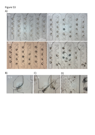
Assessment of embryo development inside the chip. A) Time-lapse images of developing zebrafish embryos collected every 24 hours. Embryos were loaded on a chip at the volumetric flow rate of 2 ml/min. Subsequently the chip was perfused at a rate of 0.4 ml/min for up to 72 hours. Only six rows are shown due to the limitation of the imaging stereoscopic system. Note the normal and very uniform development of embryos hydrodynamically immobilized on the microfluidic array; B–D) Microphotographs of hatched eletheuro-embryos at 72+ hours on a chip. Note that chip design offers the capability to recover both embryos and also swimming eletheuro-embryo stages. The recovery is best performed at the reversed flow rate when hatched stages can be collected from the inlet port. Otherwise the hydrodynamic forces will overcome the swimming behaviour and attract eletheuro-embryos back to the trapping region as denoted in D and E. |