
Fig. 7
- ID
- ZDB-FIG-120525-15
- Publication
- Narayanan et al., 2012 - Biphasic wnt8a expression is achieved through interactions of multiple regulatory inputs
- Other Figures
- All Figure Page
- Back to All Figure Page
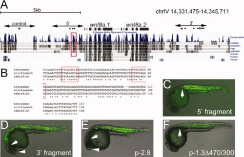
Identification of stickleback wnt8a mesoderm enhancers. A: Schematic diagram of stickleback chromosome IV spanning the wnt8a locus with the conservation tracks from the UCSC genome browser shown. The positions of the wnt8a.1 and wnt8a.2 coding regions are indicated. Control, 52, and 32 indicate the fragments tested in zebrafish transient expression assays. Dots, asterisks, and the diamond symbol above the conservation tracks indicate Ntl, FoxH1, and Zbtb4 consensus binding sequences, respectively. The red box outlines the conserved element within the margin enhancer, shown in B. B: Alignment of the conserved element. Consensus Ntl binding sequences are boxed in red. C–F: Transient expression assays in zebrafish, 24-hpf embryos, lateral view, anterior left. C: Expression from the stickleback 52 fragment extends anteriorly as far as the posterior hindbrain (arrow). Compare to the anterior limit of expression from the zebrafish -1.3Δ470/300 construct (PRR-only construct) shown in F. D: Expression from the stickleback 32 fragment extends anteriorly as far as the position of the midbrain (arrow) and some expression is observed in the telencephalon (arrowhead). Compare to transient expression from the p-2.8wnt8a.1:EGFP construct (E). |