
Fig. 2
- ID
- ZDB-FIG-120510-2
- Publication
- Song et al., 2012 - Knockdown of amyloid precursor protein in zebrafish causes defects in motor axon outgrowth
- Other Figures
- All Figure Page
- Back to All Figure Page
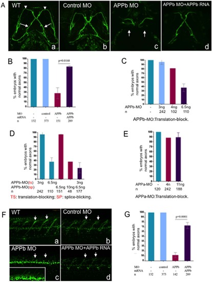
Embryos co-injected with APPb-MO and APPb mRNA rescued defective phenotypes of the Vp and VII and spinal cord nerve projections observed in APPb morphants. (A) Knockdown of APPb disrupts the projections of axons Vp and VII (3 dpf) when injected with APPb MO. Uninjected embryos and embryos injected with control MO did not show altered axonal outgrowth of neurons Vp and VII. Embryos injected with APPb-MO expressed axonal inhibition of axons Vp and VII (arrows). The defective phenotype was rescued by co-injection of APPb-MO and APPb mRNA. Ventral view; anterior, top. (B) Quantification of embryos expressing normal axonal outgrowth of neurons Vp and VII rescued through co-injection of APPb-MO (splice-block) and APPb mRNA. Uninjected embryos and embryos injected with control MO showed normal axonal growth of Vp and VII neurons. Embryos co-injected with 10 ng of APPb-MO and 350 pg APPb mRNA rescued the defected phenotype observed in the APPb morphant group. Statistical significance was established between APPb-MO embryos and co-injected embryos (p = 0.0168, p<0.05, in 2-tailed paired t test). (C) The translation-block MO of the APPb caused the identical defected phenotype on the axonal outgrowth of the Vp & VII neurons as the splice-block MO of the APPb; both were dose-dependent. (D) The morphants showed an identical defected phenotype on axonal outgrowth of Vp & VII neurons when co-injected with the translation-block MO (3 ng per embryo) of the APPb and the splice-block MO (6.5 ng per embryo) of the APPb at lower doses that did not produce a defective phenotype individually. (E) There was no defective phenotype of axonal outgrowth of Vp & VII neurons in the APPa morphants when injected with the translation-block MO against APPa. (F) APPb function is required for normal nerve outgrowth of the spinal cord. Lateral views of 3 dpf embryos (anterior is to the left, dorsal is at the top). Uninjected embryos and control embryos expressed normal motor nerve projections from the spinal cord to the myotomes (5–10 somites). Embryos injected with 10 ng of APPb-MO (splice-block) expressed severe motor neuron axon defects, including aberrant projections and decreased branching (arrows pointing at axon). Embryos co-injected with APPb-MO and APPb mRNA rescued the severe phenotype observed in the morphant group. The white rectangle in (c) is an amplification of the spinal cord neurons, which shows branching defects of the neurites in the APPb morphants. (G) Downregulation of APPb affects normal projection of motor nerves in the spinal cord. Compared with the control MO and uninjected groups, embryos injected with 10 ng APPb-MO (splice-block) showed significant axonal inhibition. Embryos co-injected with 10 ng of APPb-MO and 350 pg of APPb mRNA rescued the APPb morphant phenotype. Statistical significance was observed between morphant embryos and co-injected embryos (p = 0.0001, p<0.05 in 2-tailed paired t test). |
| Fish: | |
|---|---|
| Knockdown Reagents: | |
| Observed In: | |
| Stage: | Protruding-mouth |