
FIGURE
Fig. 4
- ID
- ZDB-FIG-120427-8
- Publication
- Chen et al., 2012 - Zebrafish agr2 is required for terminal differentiation of intestinal goblet cells
- Other Figures
- All Figure Page
- Back to All Figure Page
Fig. 4
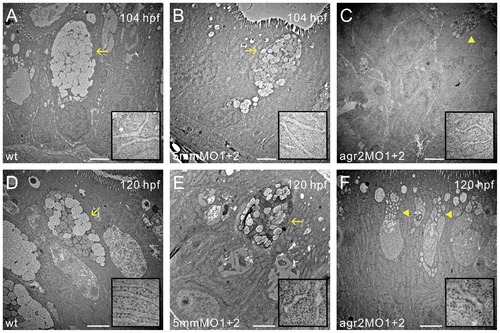
Transmission electron microscopy shows abnormal goblet cell structures in agr2 morphants. Mid-intestinal images of 104- and 120-hpf wild type (A, D), agr2–5 mmMO1 and 5 mmMO2-coinjected (B, E), and agr2 morphants (C, F) are shown. ER ultrastructure at higher magnification is shown in insets. Arrows indicate mature goblet cells and arrowheads denote immature goblet cells. Scale bars represent 2 μm. |
Expression Data
Expression Detail
Antibody Labeling
Phenotype Data
| Fish: | |
|---|---|
| Knockdown Reagents: | |
| Observed In: | |
| Stage Range: | Day 4 to Day 5 |
Phenotype Detail
Acknowledgments
This image is the copyrighted work of the attributed author or publisher, and
ZFIN has permission only to display this image to its users.
Additional permissions should be obtained from the applicable author or publisher of the image.
Full text @ PLoS One