
Fig. 4
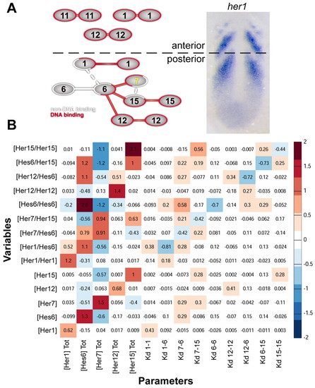
Sensitivity analysis of the dimer network. (A) Schematic of Her dimer network. DNA-binding dimers are connected in red, non-DNA-binding dimers are connected in white. Networks present in the anterior or posterior PSM are segregated and indicated on the wild-type expression of her1. Weak dimers are connected by dashed lines. (B) Summary of local sensitivity analysis of posterior PSM Her/Hes network. Input parameters (total protein concentration and Kd) are listed along the bottom and output variables (Her monomer and dimer concentrations) along the left. Sensitivity values were calculated as the fractional change in each output variable (y) for a given fractional change in the corresponding input parameter (x), to give (Δy/y)/(Δx/x), averaged across 1000 randomly sampled parameter values. Red values indicate a positive relationship, whereas blue values indicate a negative relationship between changes in parameter value and effects on Her concentration. For example, a 1% increase in total Hes6 concentration decreases Her7/Her15 by 0.56%. |