
Fig. 6
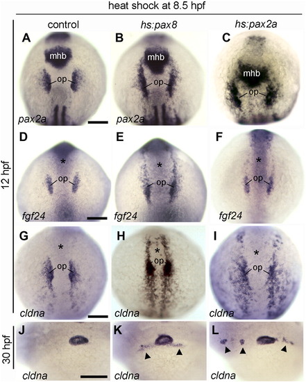
Expansion of otic markers following activation of hs:pax8 or hs:pax2a. (A–L) Dorsal views (anterior up) or lateral views (anterior to the left) of embryos heat shocked at 8.5 hpf and fixed at 12 hpf to examine expression of pax2a (A–C), fgf24 (D–F) and cldna (G–I), or embryos were fixed at 30 hpf to examine expression of cldna in the otic vesicle (J–L). Genotypes of wild-type or heterozygous transgenic embryos are indicated across the top of the figure. Expression in the otic placode (op) and midbrain–hindbrain border (mhb) is indicated. Alternatively, the position of the midbrain–hindbrain border is marked by an asterisk in (D–I). Arrowheads in K, L mark regions with ectopic expression of cldna. Scale bar, 150 μm. |
Reprinted from Developmental Biology, 364(1), Padanad, M.S., Bhat, N., Guo, B., and Riley, B.B., Conditions that influence the response to Fgf during otic placode induction, 1-10, Copyright (2012) with permission from Elsevier. Full text @ Dev. Biol.