
Fig. 5
- ID
- ZDB-FIG-120314-2
- Publication
- Trowe et al., 1996 - Mutations disrupting the ordering and topographic mapping of axons in the retinotectal projection of the zebrafish, Danio rerio
- Other Figures
- All Figure Page
- Back to All Figure Page
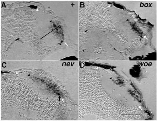
Transverse sections through the tectal lobes of a wild-type fish (A) and three mutants (B,C,D). Before sectioning, nasodorsal RGCs were labeled with DiI and the dye was photoconverted. Medial is to the left, dorsal is up. (A) A wild-type fish. The termination field of the nasodorsal axons occupies the ventral half of the tectum. Black arrows mark the ventral margin of the termination fields in all four pictures, black arrowheads the dorsal margin. The dorsal margin of the tectal lobes is labeled by white arrowheads, the ventral margin by white arrows. The black bar marks the superficialdeep axis of the tectum. (B) In box mutants, the termination field of the nasodorsal axons occupies the ventral half of the tecum, as in wild-type fish. The tectum is compressed along the superficial-deep axis. (C) In nev fish, the termination field of nasodorsal axons reaches into the dorsal side of the tecum. It is compressed along its superficial-deep axis. (D) In woe mutants, nasodorsal axons terminate in two separate fields posteroventrally and posterodorsally in the tectum. The dorsal field is oriented more towards the center of the neuropil than towards the dorsal margin. Scale bars, 50 µm. |
| Fish: | |
|---|---|
| Observed In: | |
| Stage: | Day 5 |